2023 Case Studies – Derm In-Review

June 2023 Case Study

By 2023 Case Studies

June 2023 Case Study

by Adrianna Gonzalez, MD

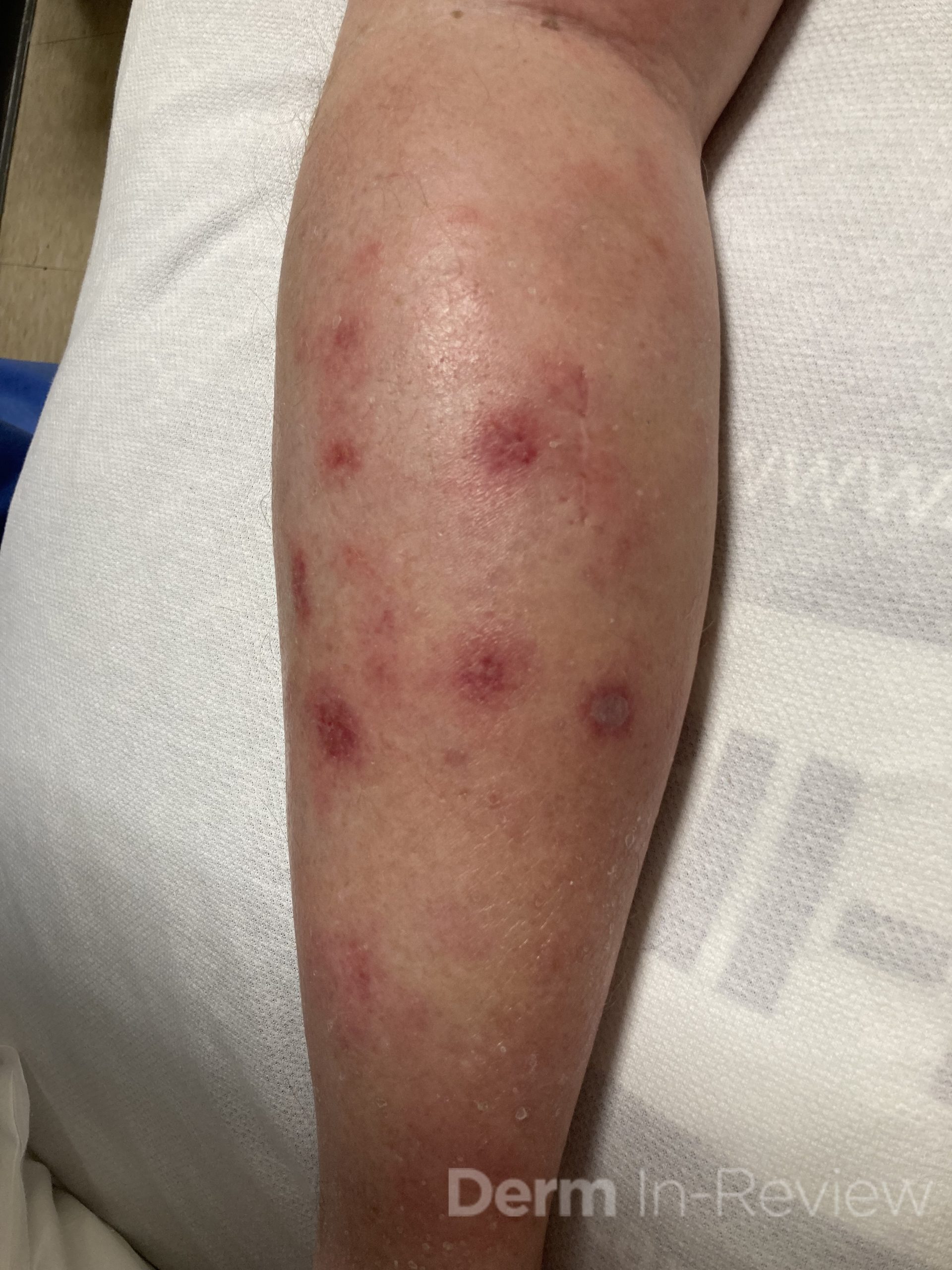
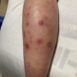
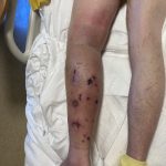
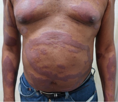
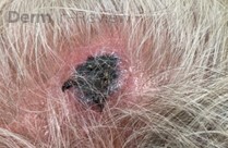
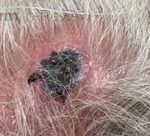
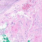
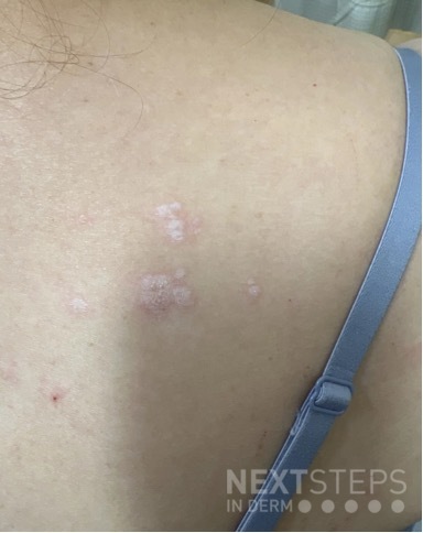
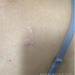
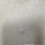
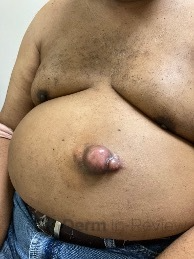
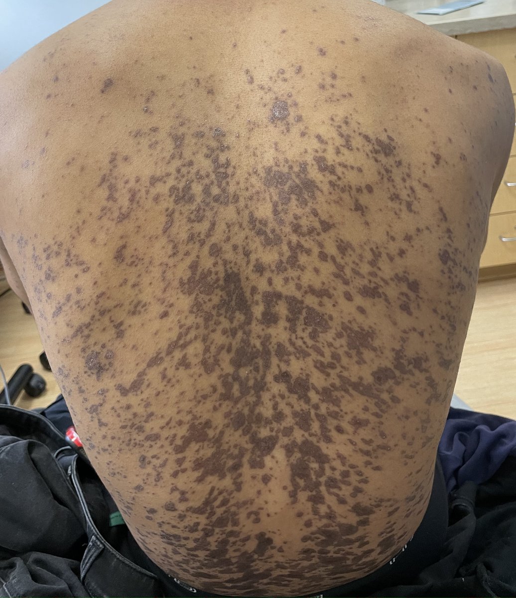
A 29-year-old male with no significant past medical history presented with complaints of a generalized rash which began two months prior. He stated the eruption began as a few lesions in the trunk that progressively grew in number and generalized to involve upper and lower extremities, buttocks, and genitals. He stated rash was associated with significant itch. Of note, he stated he had taken a new medication several weeks prior to the onset of his rash. Findings on clinical examination can be seen in Figure 1. A 4mm punch biopsy of a lesion on his mid back was performed. Histopathological examination revealed orthokeratosis, hypergranulosis, acanthosis with sawtoothing rete ridge pattern, dyskeratotic keratinocytes and a band-like lymphocytic infiltrate.

Which of the following statements is false regarding this patient’s condition?

A.) The oral mucosa is the most commonly involved body site in this entity
B.) On histology, dyskeratotic keratinocytes are classically found at the upper level of the epidermis
C.) Direct immunofluorescence may be positive for “shaggy” fibrinogen deposits along the dermoepidermal junction
D.) Flares may be triggered by certain contact allergens including copper
E.) The majority of cases will remit within 2 years


Correct Answer: B.) On histology, dyskeratotic keratinocytes are classically found at the upper level of the epidermis

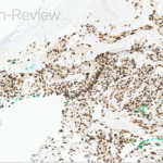
The statement in answer choice B is FALSE, which makes this the correct answer. The clinical and histopathological findings in this case are consistent with lichen planus (LP). On histology, LP is characterized by compact orthokeratosis, wedge-shaped hypergranulosis, sawtooth pattern of rete ridges, vacuolar degeneration of the basal layer, dyskeratotic keratinocytes (Civatte bodies), and a band-like lymphocytic infiltrate at the dermoepidermal junction (DEJ). Dyskeratotic keratinocytes are classically present at the lower levels of the epidermis and at the superficial papillary dermis near the basal layer, the site of destruction.1 Dyskeratotic keratinocytes can be seen in the suprabasilar, upper epidermal layer in erythema multiforme, Stevens-Johnson Syndrome and toxic epidermal necrolysis.2 Direct immunofluorescence (DIF) demonstrates “shaggy” deposition of fibrinogen along the basement membrane zone, as well as staining of colloid bodies with one or more of IgM, IgG, IgA and C3  (answer C).

Lichen planus is an inflammatory disorder of the skin, mucosa, hair and nails. Although the incidence appears to vary throughout geographical areas, the oral mucosa has been found to be the most commonly involved body site in most study populations (answer A).1,3 Oral lesions can be observed in over 50% of patients with LP, and can often be the only site of involvement.2 A recent epidemiological study estimated that the global pooled prevalence of oral LP was 0.89% among the general population and 0.98% among clinical patients.4 Cutaneous LP is thought to affect < 1% of the population worldwide and commonly presents as pruritic, purple, polygonal, flat-topped papules commonly involving the extremities and lower back.5 There are numerous variants of cutaneous LP, including actinic, annular, atrophic, bullous, drug-induced, genital, hypertrophic, inverse, linear, LP pigmentosus and lichen planopilaris. LP may also involve the nails and present with longitudinal ridging, trachyonychia, thinning, dorsal pterygium and even 20 nail dystrophy. While its pathophysiology has not been fully elucidated, LP is thought to be caused by an altered cellular immune response, in which cytotoxic CD8+ T cells attack basal keratinocytes. LP has been associated with various triggers, including infections such as hepatitis C virus (more common in oral erosive LP), medications, contact allergens, vaccinations (hepatitis B virus and influenza) and genetics. Implicated contact allergens include gold, mercury amalgam and copper (answer D).1  Most cases of LP self-resolve within 1-2 years (answer E). Oral LP (especially oral erosive LP), nail LP and hypertrophic LP tend to be persistent and recalcitrant to treatment.

References

  1. Boch K, Langan EA, Kridin K, Zillikens D, Ludwig RJ, Bieber K. Lichen Planus. Front Med (Lausanne). 2021;8:737813.
  2. Bolognia J, Jorizzo, J. L., & Schaffer, J. Dermatology. Fourth Edition ed. Philadelphia: Elsevier Saunders; 2012.
  3. Arnold DL, Krishnamurthy K. Lichen Planus. In: StatPearls. Treasure Island (FL)2022.
  4. Li C, Tang X, Zheng X, et al. Global Prevalence and Incidence Estimates of Oral Lichen Planus: A Systematic Review and Meta-analysis. JAMA Dermatol. 2020;156(2):172-181.
  5. Tziotzios C, Lee JYW, Brier T, et al. Lichen planus and lichenoid dermatoses: Clinical overview and molecular basis. J Am Acad Dermatol. 2018;79(5):789-804.

May 2023 Case Study

By 2023 Case Studies

May 2023 Case Study

by Emily Murphy, MD1, Karl Saardi, MD1

  1. Department of Dermatology, George Washington University School of Medicine and Health Sciences

A 65-year-old male with a past medical history of HIV (CD4 count 250, undetectable viral load) and chronic congestion presented with a chief complaint of recurrent cellulitis of the left lower leg. The redness and swelling persisted for 3 months despite antibiotics. On presentation, he had circumferential edema and induration of the left lower leg with erythematous, tender nodules and early bullae (figure 1). A week later, his disease progressed with ulceration and necrosis of these tender nodules (figure 2). A punch biopsy was done, which showed an atypical lymphoid infiltrate in the dermis and subcutaneous tissue with an angiocentric pattern, composed of large, pleomorphic cells. Extensive necrosis was present with karyorrhetic debris. Immunostains were positive for T cells (CD2), natural killer cells (CD56), and cytotoxicity (granzyme B). A biopsy of the nasal septum showed similar findings. The patient was diagnosed with extranodal NK/T cell lymphoma, nasal type.

Which virus is part of the diagnostic criteria for this lymphoma?

A.) Human herpesvirus-8
B.) Polyomavirus
C.) Human immunodeficiency virus
D.) Ebstein Bar Virus
E.) Cytomegalovirus

Correct Answer: D) Ebstein Bar Virus

Extranodal NK/T cell lymphoma, nasal type is an aggressive, non-Hodgkin lymphoma that has four criteria according to the World Health Organization: vascular damage, prominent necrosis, a cytotoxic phenotype, and association with the Ebstein bar virus (EBV, answer D).1,2 EBV positivity is seen in the both the skin and serum. Pathologically, the atypical lymphocytes are positive for cytotoxic markers (granzyme B, peforin), natural killer cell markers (CD2), T cell markers (cytoplasmic CD3), and EBV.1 Extranodal NK/T cell lymphoma is rare in the United States, but its incidence is increasing. It is more common in East Asia and Latin America.1

This lymphoma typically affects the upper aerodigestive tract, causing congestion, epistaxis, or necrotizing lesions of the nose or hard palate.1 Other sites can be involved, including the extranasal skin, gastrointestinal tract, testes, or lungs.1,3,4 On the skin, either a generalized morbilliform eruption or purpuric nodules with or without ulceration can be seen.3 Hemophagocytic lymphohistiocytosis can occur, but is rare, occurring in only 3% of patients.1

NK/T cell lymphoma has an increased incidence in patients with HIV; a study of 93 patients with this lymphoma found that it occurred at a 15 times higher incidence in patients with HIV compared to the general population.5 However, HIV (choice C) is not part of the diagnostic criteria like EBV. Human herpesvirus-8 (Kaposi sarcoma), Polyomavirus (Merkel Cell Carcinoma), and cytomegalovirus are not associated with Extranodal NK/T cell lymphoma (choices A, B, E).

Treatment is determined by the Ann Arbor Staging and various prognostic tools, like the PINK-E system (age > 60 years, Stage III/IV, nonnasal primary localization, distant lymph node involvement, and detectable plasma EBV DNA).6 Depending on the stage and prognosis, treatment is typically with an asparaginase-based regimen called SMILE (dexamethasone, methotrexate, ifosfamide, L-asparaginase, and etoposide).1,6,7 NK/T cell lymphoma cells are unable to synthesize the amino acid, asparagine, and as this amino acid is depleted with asparaginase, protein synthesis is disrupted and the cells ultimately undergo apoptosis.6 Chemotherapy is typically followed by involved field radiation therapy.1  Various immunotherapies are currently being studied for patients with recalcitrant or relapsed disease as well, including programmed death 1 inhibitors, JAK 3 inhibitors, or anti-CD30 therapies.8 Response to therapy can be monitored with EBV DNA titers, which should decrease with treatment.1

References

  1. Haverkos BM, Pan Z, Gru AA, et al. Extranodal NK/T Cell Lymphoma, Nasal Type (ENKTL-NT): An Update on Epidemiology, Clinical Presentation, and Natural History in North American and European Cases. Curr Hematol Malig Rep. 2016;11(6):514-527. doi:10.1007/s11899-016-0355-9
  2. Willemze R, Cerroni L, Kempf W, et al. The 2018 update of the WHO-EORTC classification for primary cutaneous lymphomas. Blood. 2019;133(16):1703-1714. doi:10.1182/blood-2018-11-881268
  3. Orlowski GM, Tan AJ, Evan-Browning E, Scharf MJ. Primary cutaneous nasal-type NK/T-cell lymphoma presenting as purpuric nodules on the lower leg. JAAD Case Rep. 2020;6(10):1075-1078. doi:10.1016/j.jdcr.2020.08.007
  4. Rahal A, Reddy PS, Alvares C. Extranodal NK/T-Cell Lymphoma, Nasal Type, Presenting as a Breast Mass. Cureus. 7(12):e408. doi:10.7759/cureus.408
  5. Castillo J, Pantanowitz L. HIV-Associated NK/T-Cell Lymphomas: A Review of 93 Cases. Blood. 2007;110(11):3457-3457. doi:10.1182/blood.V110.11.3457.3457
  6. van Doesum JA, Niezink AGH, Huls GA, Beijert M, Diepstra A, van Meerten T. Extranodal Natural Killer/T-cell Lymphoma, Nasal Type: Diagnosis and Treatment. Hemasphere. 2021;5(2):e523. doi:10.1097/HS9.0000000000000523
  7. Yamaguchi M, Kwong YL, Kim WS, et al. Phase II study of SMILE chemotherapy for newly diagnosed stage IV, relapsed, or refractory extranodal natural killer (NK)/T-cell lymphoma, nasal type: the NK-Cell Tumor Study Group study. J Clin Oncol. 2011;29(33):4410-4416. doi:10.1200/JCO.2011.35.6287
  8. Lv K, Li X, Yu H, Chen X, Zhang M, Wu X. Selection of new immunotherapy targets for NK/T cell lymphoma. Am J Transl Res. 2020;12(11):7034-7047.

April 2023 Case Study

By 2023 Case Studies

April 2023 Case Study

by Blair Allais, MD

A 63-year-old male presented to clinic with a 2-year history of asymptomatic annular hypopigmented plaques on the trunk and extremities that eventually became erythematous to violaceous. He had been prescribed topical steroids by an outside practitioner without improvement in the lesions. Biopsy performed on one of the plaques revealed a granulomatous reaction surrounding adnexal structures and in the papillary dermis. Fite stain revealed numerous pink to red acid-fast organisms within the granulomas ultimately rendering a diagnosis of lepromatous leprosy. The patient was started on therapy with Rifampin 600mg monthly, Minocycline 100mg monthly and Moxifloxacin 400mg monthly. Approximately 3 months after initiating treatment the patient developed new nodular skin lesions on the extensor arms and medial thighs associated with fever, myalgias, fatigue and joint pain.

What is the best course of treatment for this patient’s new presentation?

A.) Prednisone
B.) Clofazimine
C.) Thalidomide
D.) Methotrexate

Correct Answer: C) Thalidomide 

The patient described has a diagnosis of lepromatous leprosy and is experiencing a reactional state to treatment. Approximately 50% of patients with leprosy will experience a reaction after the institution of therapy. Aside from initiating antibiotic therapy, other causes of reactional states include intercurrent infections, vaccination, pregnancy, vitamin A, iodides and bromide. Reactions are an important cause of permanent nerve damage in borderline patients and are abrupt in appearance.

Two major types of reactional states have been described. Type 1 reactions due to enhancement of cell-mediated immunity with a Th1 cytokine pattern (IFN-y, TNF, IL-1ß, IL-2, IL-12). These reactions are considered to reversal reactions with “upgrading” and are seen predominantly in borderline or tuberculoid forms of leprosy. They are characterized by increased inflammation within existing skin lesions in addition to acute nerve pain or tenderness (neuritis) and loss of function. Treatment of choice for these reactions is prednisone (A) starting at a dose of 40 – 60 mg/day. Neuritis and eye lesions are urgent indications for therapy and dose and duration is determined by the clinical course of the reaction. Steroids are tapered slowly over months to years once the reaction is controlled. In some cases, clofazimine (B) may be added as it has some activity against the type 1 reaction, and may be added in doses of up to 300 mg/day as tolerated.

Type 2 reactions are due to excessive humoral immunity with a Th2 cytokine pattern (IL-4, IL-10) and characterized by formation of immune complexes. They are seen predominately in patients with lepromatous and borderline lepromatous forms of leprosy, especially in patients with a high bacterial load who are undergoing treatment. Multisystem involvement is seen with systemic symptoms including nodular skin lesions, fever, myalgias, malaise, severe joint swelling and pain, iridocyclitis, lymphadenitis, hepatosplenomegaly, orchitis and glomerulonephritis. The intensity of the reaction can vary from mild to severe and can last from a few days to weeks, months, or even years. Pathology is characterized by cutaneous and systemic small vessel vasculitis, and treatment of choice is Thalidomide (C). According to the newly developed U.S. regimen for treatment for lepromatous leprosy, methotrexate (D) 15mg weekly can be prescribed to prevent reactions at the time of initiating multi drug treatment.


References

  1. Ramos-e-Silva M, Ribeiro de Castro MC. Chapter 75: Mycobacterial Infections. 4th Ed. Philadelphia, PA: Elsevier; 2018.
  2. James WD et al. Chapter 17: Hansen Disease. Andrews’ Diseases of the Skin. 13th Philadelphia, PA: Elsevier; 2020.
  3. Bennett JE et al. Principles and Practice of Infectious Diseases. 6th edit.P.2887-2896

March 2023 Case Study

By 2023 Case Studies

March 2023 Case Study

by Emily Murphy, MD1

  1. Department of Dermatology, George Washington University School of Medicine and Health Sciences

A 70-year-old healthy male presented with a chronic wound on his scalp that started 4 months ago. On examination, a 2.5-centimeter circular, ulcerated violaceous plaque with hemorrhagic crusting was present on the scalp (figure 1). A punch biopsy of the raised border was done (figure 2a, hematoxylin-eosin staining). The atypical cells stained positively for erythroblast transformation specific related gene (ERG) and CD31.

Based on the clinical presentation and biopsy findings, which two patients would be least likely to develop the same malignancy as the above patient?

A.) 80-year-old male with a plaque on the cheek
B.) 35-year-old male with a tumor that also stained positively for HHV-8
C.) 65-year-old male exposed to vinyl chloride for many years in his occupation
D.) 50-year-old woman with a lower limb plaque after treatment of a uterine carcinoma 10 years prior
E.) 55-year-old woman with a plaque on the scalp who reports chronic sun exposure and many blistering sunburns
F.) 40-year-old woman who had radiation for breast cancer 5 years ago


Correct Answer: B, E

Given the biopsy revealed proliferation of atypical and inter-anastomosing vascular channels along with increased mitotic figures, and that these atypical cells were positive for ERG and CD31, the patient was diagnosed with angiosarcoma, a rare soft tissue sarcoma, with only about 200 reported cases in the United States per year.1 These tumors present on the skin as a purpura-like or violaceous papules to plaques, that are often multifocal.2 As the tumors grow, ulceration and hemorrhage often occur, as was seen in this case.2

The cutaneous form of angiosarcoma most often occurs spontaneously, typically in elderly men on the head and neck (choice A).2 The scalp is a particularly common site, as in our patient, and is also associated with a poor prognosis.3,4 Angiosarcomas can also occur secondary to radiation (choice F) or chronic lymphedema (choice D), which is called Stewart-Treves syndrome.1,2 Stewart-Treves can be seen after lymph node dissection in setting of breast cancer (upper limb) or uterine cancer (lower limb, choice D).1 In cases of radiation or lymphedema, the tumor often develops at least 5 years later.1,2 Angiosarcoma can also be seen with chronic exposure to various toxins including vinyl chloride, arsenic, thorium dioxide, and anabolic steroids (choice C).2 Unlike Kaposi sarcoma, HHV-8 is not associated with angiosarcoma (choice B).6 Further, in contrast to many other cutaneous cancers, sun exposure is not associated with angiosarcomas (choice E).7

Prior to treatment, an MRI of the primary lesion should be done to evaluate the extent of disease, as well as a CT chest to rule of pulmonary disease, which is the most common site of metastasis.1,2 A PET scan may also be done to exclude other sites of metastasis.2 Given the rarity of these tumors, standard treatment algorithms do not exist. For local disease, wide local excision is done, typically followed by surgical site radiation of at least 50 gray.1 For unresectable or metastatic disease, chemotherapy, often paclitaxel, is used in combination with radiation.1 Clinical trials are currently examining the impact of immunotherapy on angiosarcoma as well. Despite treatment, these tumors often recur, leading to a 5-year survival less than 40%.1


References

  1. Fujisawa Y, Yoshino K, Fujimura T, et al. Cutaneous Angiosarcoma: The Possibility of New Treatment Options Especially for Patients with Large Primary Tumor. Front Oncol. 2018;8:46. doi:10.3389/fonc.2018.00046
  2. Young RJ, Brown NJ, Reed MW, Hughes D, Woll PJ. Angiosarcoma. Lancet Oncol. 2010;11(10):983-991. doi:10.1016/S1470-2045(10)70023-1
  3. Ramakrishnan N, Mokhtari R, Charville GW, Bui N, Ganjoo K. Cutaneous Angiosarcoma of the Head and Neck-A Retrospective Analysis of 47 Patients. Cancers (Basel). 2022;14(15):3841. doi:10.3390/cancers14153841
  4. Shin JY, Roh SG, Lee NH, Yang KM. Predisposing factors for poor prognosis of angiosarcoma of the scalp and face: Systematic review and meta-analysis. Head Neck. 2017;39(2):380-386. doi:10.1002/hed.24554
  5. Jennings TA, Peterson L, Axiotis CA, Friedlaender GE, Cooke RA, Rosai J. Angiosarcoma associated with foreign body material. A report of three cases. Cancer. 1988;62(11):2436-2444. doi:10.1002/1097-0142(19881201)62:11<2436::aid-cncr2820621132>3.0.co;2-j
  6. Schmid H, Zietz C. Human herpesvirus 8 and angiosarcoma: analysis of 40 cases and review of the literature. Pathology. 2005;37(4):284-287. doi:10.1080/00313020500169495
  7. North P. Vascular Neoplasms and Neoplastic-Like Proliferations. In: Dermatology. Fourth Edition. Elsevier; :2020-2049. Accessed February 18, 2023. https://www-clinicalkey-com.proxygw.wrlc.org/#!/content/book/3-s2.0-B9780702062759001148?scrollTo=%23top

February 2023 Case Study

By 2023 Case Studies

February 2023 Case Study

by Alexis E. Carrington, MD

A 52 year old female with a past medical history of vulvar lichen sclerosus presents to the clinic for a rash on the back and legs for 8 months. She reports the rash is itchy and the areas on the back are worse during the summer months and with sweating. She is using Aquaphor ointment and a topical antibiotic cream with no improvement. She denies any family history of similar findings or exposure to new personal or household items. Punch biopsy of a lesion on the back is consistent with lichen sclerosus.

Which of the following is true about lichen sclerosus?

A.) It is associated with HLA-DQ2
B.) It is associated with anti-extracellular matrix protein 1 antibodies
C.) Genital lesions can be associated with development of BCC
D.) Treatment of extragenital disease involves the use of a low-potency topical steroid


Extragenital lichen sclerosus typically favors the neck, shoulders, trunk, proximal extremities, flexor wrists and sites of trauma. It is associated with xerosis and mild pruritus, but is usually asymptomatic. The lesions start as white, shiny, polygonal papules that coalesce into plaques and evolve into atrophic patches and plaques. Lesions may also affect the skin around the eye and other areas of the face.

Lichen sclerosus is usually a clinical diagnosis but a biopsy is reasonable to confirm diagnosis. Topical steroids are the gold standard for this condition. Patients should be referred to a dermatologist and gynecologist or urologist for management of genitourinary complications.

Correct answer: B.) It is associated with anti-extracellular matrix protein 1 antibodies

Answer A is associated with Dermatitis Herpetiformis, whereas lichen sclerosus is associated with HLA-DQ7. There is an increased risk of genital squamous cell carcinoma in both men and women with lichen sclerosus, not basal cell carcinoma (Answer C). Treatment of extragenital disease involves the use of high potency topical steroids, rather than low-potency (Answer D).


References

  1. Bolognia, Jean Dermatology volume 2. Mosby, 2018.
  2. Fistarol SK, Itin Diagnosis and treatment of lichen sclerosus: an update. Am J Clin Dermatol. 2013 Feb;14(1):27-47. PubMed ID: 23329078
  3. Lewis FM, Tatnall FM, Velangi SS, et al. British Association of Dermatologists guidelines for the management of lichen sclerosus, Br J Dermatol. 2018 Apr;178(4):839-853. PubMed ID: 29313888

January 2023 Case Study

By 2023 Case Studies

January 2023 Case Study

by Adam Rosenfeld, MD

61-year-old male with history of HTN, DM, HLD who presented for a new growth on his mid abdomen. He noted a history of a trauma to the area with a metal blade 20 years prior while in active duty, with the area initially healing with a raised scar. He endorsed significant increase in size of the area over the last year, but it had been slowly growing for several years. The patient had been told this was a keloid. He denies any significant pain or associated symptoms. On examination, there was a large approximate 4x5cm protuberant skin colored to pink mass on the left mid abdomen (figure 1). A punch biopsy was obtained of this tumor demonstrating a dense dermal infiltrate of spindle cells (figure 2).

A diagnosis of a malignant dermal proliferation was made, often mimicking the diagnosis the patient had been told before. What stain was confirmatory in making this diagnosis?

A.) CD207+
B.) Factor XIIIA+
C.) S100+
D.) CD34+
E.) SMA+

Correct answer: D.) CD34+

This multi-loculated tumor is suspicious for dermatofibrosarcoma protuberans, or DFSP which classically stains diffusely positive for CD34. CD34 is a marker for vascular endothelium and hematopoietic progenitor cells4. It is positive in DFSP and negative in dermatofibromas. Dermatofibrosarcoma protuberans is a fibroblast derived intermediate-grade soft tissue sarcoma that can be mistaken for a keloid3. It is often slow growing and asymptomatic. It can present as a bulky multi-loculated tumor as in our patient but can also have much more innocent appearances. Its pathophysiology is unclear, but some have proposed prior injury to the skin as a triggering event1. Evidence does support these tumors arising from fibroblastic, histiocytic or neuroectoderm tissue although a fibroblastic origin seems most likely. The growth of the tumor is driven in most cases by a translocation of chromosome 17 and 22 forming the t(17,22) COLA1A-PDGFG fusion protein1. On histopathology, a dense dermal infiltrate of spindle cells is seen infiltrating into the deeper fat causing the “honey combing” appearance of the adipose tissue4. These cells stain diffusely positive for CD34+, while staining weakly or not at all for Factor XIIIA+ (answer choice B), the stain pathognomonic for dermatofibromas4. Importantly, it is crucial to rule out other spindle cell neoplasms including a spindle cell melanoma which would stain positive for S100 (answer choice C)4. While DFSPs are malignant, they typically do not demonstrate significant atypia histologically. Fibrosarcomatous change represents a subset of DFSPs that demonstrate much more atypical features, indicating tumor progression3. The differential diagnosis of DFSP includes but is not limited to a keloid, dermatofibroma, spindle cell melanoma, squamous cell carcinoma, Kaposi sarcoma.

The standard of care for treatment is surgical excision with every effort to achieve clear surgical margins both peripherally and deep2. Although wide local excision can be performed, Mohs Micrographic Surgery (MMS) is now recommended for these tumors. Given that it demonstrates unpredictable microscopic, tentacle like extensions, the directionality afforded by MMS makes it uniquely fit for DFSPs3. No set margins exist but wide local excision typically includes 2-4cm. MMS initial margins vary but the literature suggest 0.5-1.3cm3. Unresectable or metastatic tumors can be treated with radiation or Imatinib, a tyrosine kinase inhibitor5,7. NCCN guidelines do not recommended extensive systemic work up of these patients unless there are red flags for metastasis on exam or through history. Imaging with CT or MRI can be performed to assess for tumor depth but is based on a case-by-case basis8. These tumors have a low risk of metastasis (1-4%) but a high chance of local recurrence. Surveillance includes observation of primary site every 6 months for the first 5 years with yearly exams following3. The overall prognosis of DFSPS is good, with a 10-year survival rate of 99.1%3.

Incorrect stains in answer choices4

CD 207 (choice A) – surrogate marker for presence of Birbeck granules in Langerhans cells

Factor XIIIA (choice B) – stains dermal dendritic cells, positive in dermatofibroma and negative in DFSP

S100 (choice C) – stains melanocytes, Langerhans cells, sweat glands, nerves, Schwann cells, myoepithelial cells, fat, muscle and chondrocytes

SMA (choice E) – positive in smooth muscle, including vascular smooth muscle; negative in skeletal muscle; expressed in myofibroblasts and myoepithelial cells


References

  1. Bolognia, J., Schaffer, J., & Cerroni, L. (2018). Dermatology (Fourth edition.). Philadelphia,Pa: Elsevier.
  2. Dermatofibrosarcoma protuberans – statpearls – NCBI bookshelf. Dermatofibrosarcoma Protuberans. (n.d.). Retrieved August 11, 2022, from https://www.ncbi.nlm.nih.gov/books/NBK513305/
  3. Hao X;Billings SD;Wu F;Stultz TW;Procop GW;Mirkin G;Vidimos AT; (n.d.). Dermatofibrosarcoma protuberans: Update on the diagnosis and treatment. Journal of clinical medicine. Retrieved August 11, 2022, from https://pubmed.ncbi.nlm.nih.gov/32516921/
  4. Elston, D. M. (2019). Dermatopathology. Elsevier.
  5. Cristian Navarrete-Dechent, M. D. (2019, March 1). Imatinib treatment for locally advanced or metastatic dermatofibrosarcoma protuberans. JAMA Dermatology. Retrieved August 11, 2022, from https://jamanetwork.com/journals/jamadermatology/article-abstract/2719525
  6. Raman K Madan, M. D. (2021, July 15). Dermatofibrosarcoma protuberans. Background, Pathophysiology, Etiology. Retrieved August 11, 2022, from https://emedicine.medscape.com/article/1100203-overview
  7. Ugurel, S., Mentzel, T., Utikal, J., Helmbold, P., Mohr, P., Pföhler, C., Schiller, M., Hauschild, A., Hein, R., Kämpgen, E., Kellner, I., Leverkus, M., Becker, J. C., Ströbel, P., & Schadendorf, D. (2014, January 16). Neoadjuvant imatinib in advanced primary or locally recurrent dermatofibrosarcoma protuberans: A multicenter phase II decog trial with long-term follow-up. American Association for Cancer Research. Retrieved August 15, 2022, from https://aacrjournals.org/clincancerres/article/20/2/499/78307/Neoadjuvant-Imatinib-in-Advanced-Primary-or
  8. Bichakjian, C. K., Olencki, T., Alam, M., Andersen, J. S., Berg, D., Bowen, G. M., Cheney, R. T., Daniels, G. A., Glass, L. F., Grekin, R. C., Grossman, K., Ho, A. L., Lewis, K. D., Lydiatt, D. D., Morrison, W. H., Nehal, K. S., Nelson, K. C., Nghiem, P., Perlis, C. S., … Ho, M. (2014, June 1).Dermatofibrosarcoma protuberans, version 1.2014. JNCCN. Retrieved January 9, 2023, from https://jnccn.org/view/journals/jnccn/12/6/article-p863.xml